Phineas Gage and the Iron Rod Incident
Case that revolutionised our approach to studying human behaviour
Outline
NeuraFind’s first pick for a case study is Phineas Gage and the iron rod accident. This may be one some of you have already heard about but its an important one. We’re starting with a gory and bizarre case study that revolutionised the way we investigate behaviour in humans.
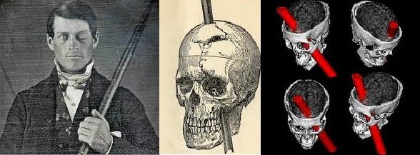
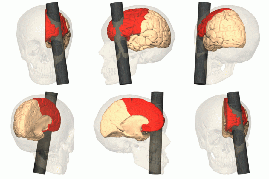
Phineas was a 25-year old Railway Construction Supervisor working in Vermont at the Rutland and Burland Railroad Site. On September 13th, 1848, he was preparing powder charge to blast rocks when an unfortunate explosion led to a 2.5cm wide and 1 m long iron rod getting lodged into his skull. It entered through his left cheek, destroyed his eye and the frontal lobe of his brain. It left the top of his skull. He had lost consciousness almost immediately.
Amazingly though, he was able to regain consciousness, talk and walk to the hospital himself. At the hospital, his physician, John M Harlow treated him. In his “Passage of an Iron Rod Through the Head”, he logs Gage’s overall treatment until recovery. In it, he describes the scene in a very gory manner - he mentions that “the bed on which he was laid, were literally one gore of blood.”. He then goes onto describing the wound - “the fragments of bone were uplifted and the brain protruding”.
Amazingly though, he gained consciousness in a few minutes of the accident.
John M Harlow, an American Physician treated him. In his article, “Passage of an Iron Rod Through the Head”, he logs the details of Gage’s overall treatment.
Harlow describes the scene as “the bed on which he was laid, were literally one gore of blood.” And that “the fragments of bone were uplifted and the brain protruding”.
According to Harlow, Gage’s pulse remained 60-100 (regular) throughout the treatment. The variations were quite soft and nothing that was a cause for concern. There was quite a bit of internal and external haemorrhaging (bleeding) in the initial stages - at some point he was bleeding into his stomach. After the lobotomy, the haemorrhaging had slowed down a lot but did continue a little bit. Gage was able to recognize his friends and seemed rational. He also vomited quite a few times after the surgery. Nausea and vomiting it quite common after brain surgeries. This is because of the altered pressure in the head, which may cause a queasy sensation.
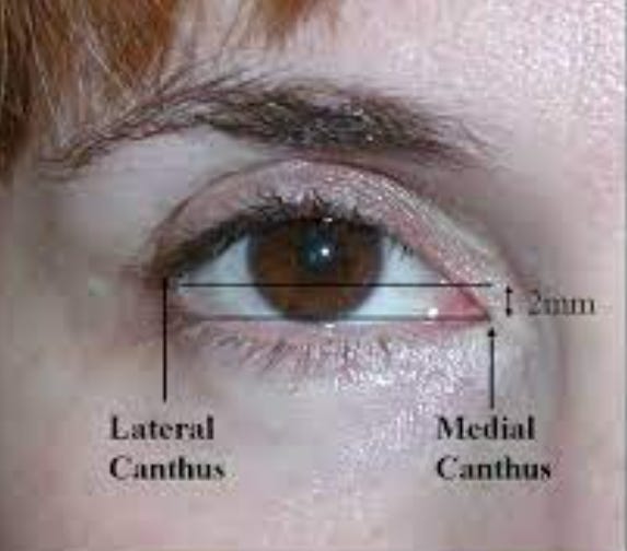
On the 3rd day after the incident, a fungus was found growing in his external canthus of his left eye. Risk of infections and fungus is common after any surgery. And since Gage’s case involved foreign objects (rod), this increased the chances of an infection.
Gage had a couple of sleepless and restless nights. He threw hands and feet and tried to get out of bed.
In just two months, Gage was up and walking. He had recovered almost completely. He had lost his eye but that didn’t stop him from living on.
Here’s the catch though – Gage’s personality had changed.
He had become arrogant, extravagant, dishonest, ill-mannered, impulsive and a lot more. There have been plenty allegations of sexual indecency, hedonistic behaviour, violence and many more. Although these allegations may not be true, one thing was for sure that he had changed.
His friends and family claimed
“Gage was not Gage anymore”.
Damages
Firstly, his hands and fore arms were both deeply burned nearly to the elbows due to the explosion. These were dressed. The rod had gone through the left eye and destroyed it entirely.
Muscular and Skeletal Damages
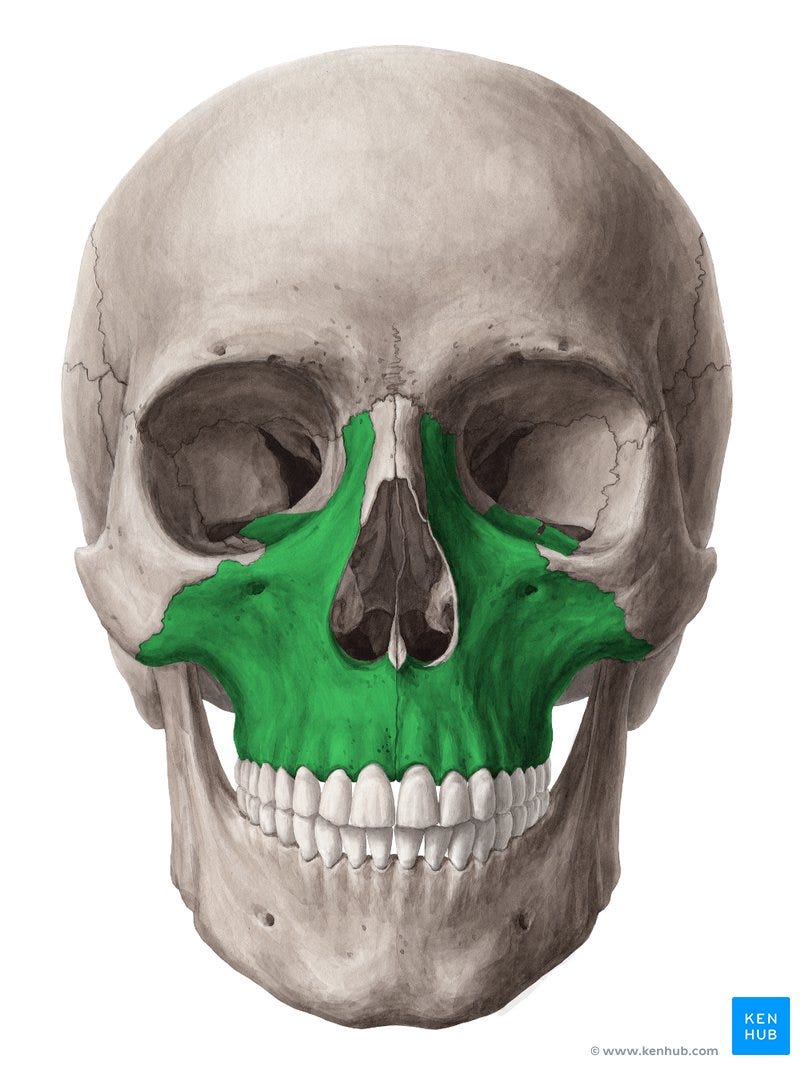
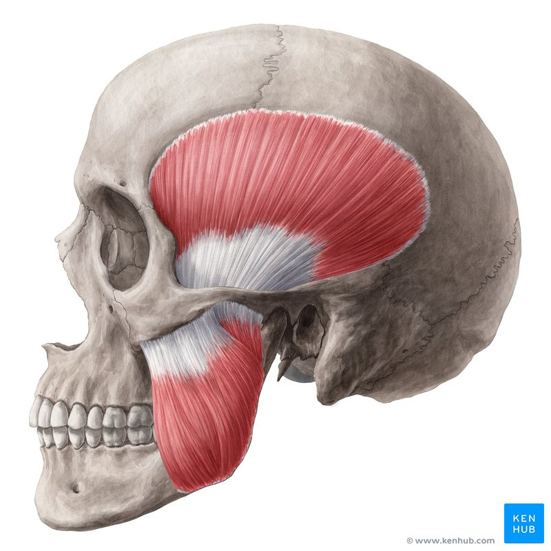
The rod penetrated the skull through the inferior maxillary bone. The maxillary bone is the bone that makes up the upper jaw as shown in the diagram below. The rod contacted the inferior part of this bone.
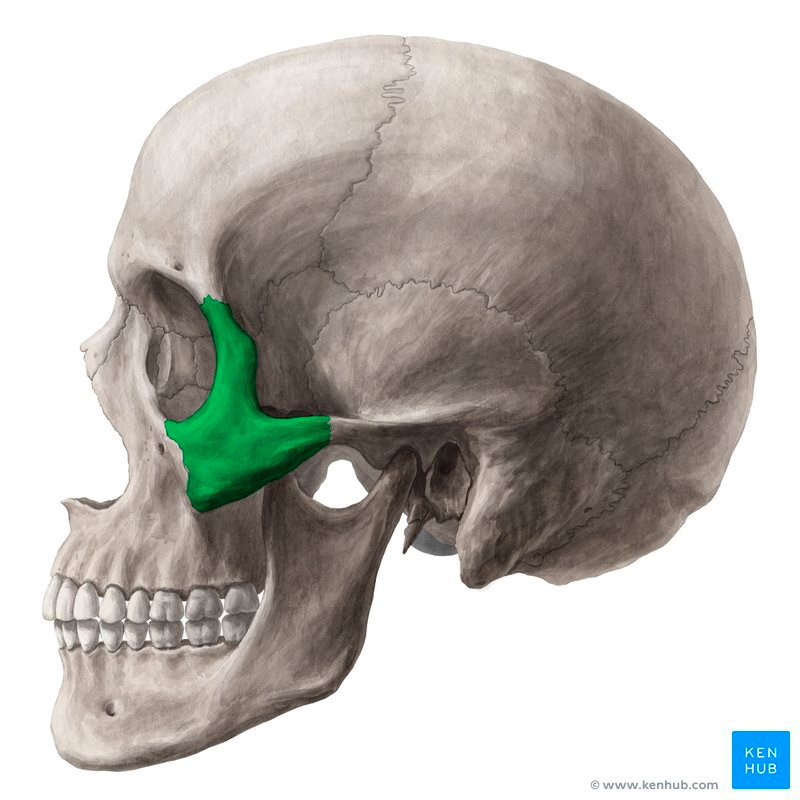
Essentially, it placed itself right inside the zygomatic arch, which is shown below.
In the process, it tore through the masseter and temporal muscles, which lie next to the zygomatic arch.
It left the skull through the parietal and frontal bones. Overall, it looked as following.
Brain Damage
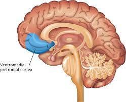
Harlow claimed that all the damage was done entirely to the anterior left lobe of the cerebrum. However, the ventromedial (the bottom middle) region of both the frontal lobes were damaged. The parts of the brain that are responsible for speech and motor functions, motor cortex and broca’s area, were spared. This is why Gage was able to speak and move as normal.
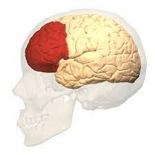
It was the prefrontal cortex that suffered the most damage. This is located in the ventromedial region of the frontal lobe and shown in the images below
Neuroscientific Analysis
Phineas Gage’s case revolutionised the way we investigate behaviour. It was because of this case that the link between behaviour and the brain parts.
In extremely simple terms, it was the to his prefrontal cortex that changed him. The prefrontal cortex is responsible for rational cognitive functions – it is known help control impulses aka stopping us from acting out. It is the voice of reason. In fact modern neuroscience has noticed a shrinkage in the prefrontal cortex of serial killers. This was the theory that was most commonly known.
However, in 2012, Van Horn et al, used Computed Tomography (CT) Diffusion Weighted Imaging (DWI) and Magnetic Resonance Imaging (MRI), they assessed the extent of cortical gray matter (GM) and white matter (WM) damage.
Alright, I know, many terms, let me break this down.
Computed Tomography (CT)
These use series of X-ray images from different angles. This is processed in a computerized system to produce cross-sectional slices of the target parts. To understand this, you need to understand how X-rays work.
X-ray radiation is directed towards the target spot. Areas that are not penetrable absorb the rays which shows up on the film as white. The spaces where the energy passes right through are the darker areas.
This can be used to find out any irregularities in the brain.
Magnetic Resonance Imaging (MRI)
These use magnetic and radio waves to map the internal parts of the body.
Diffusion Weighted Imaging (DWI)
This is one that is very popular for white matter mapping. This detects the difference in the Brownian motion of the water molecules within different material. By looking for these contrasts, it is able to map the white matter.
Van Horn was able to use these techniques along with data about Gage’s skull through his CTs to accurately model the affected areas. They computationally simulated the rod penetrating his skull.
Firstly, they noticed that there in fact was quite a lot of damage to left frontal cortex. The impact of this affected the connections to other parts of the brain too. Essentially, there was a significant connectivity loss in the WM between the frontal regions and the rest of the brain.
This led to a more holistic approach to brain localization - Unlike what Harlow Suggested, personality has to do with more than just left frontal lobe.


